Coronary Angiography
What is a coronary angiogram?
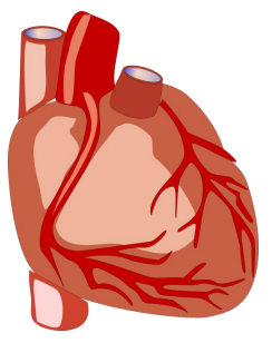
A coronary angiogram (also often called an angiogram or cardiac catheterisation) is a test that is used to look at the main arteries (coronary arteries), which supply the heart with fresh blood and oxygen.
A coronary angiogram is a special type of x-ray that will show your cardiologist the exact location and condition of any narrowed areas in your coronary arteries.
Why do I need one?
A coronary angiogram can be used to help diagnose heart conditions and help plan future treatments. You may need this test if your cardiologist thinks that you have a problem with one or more of your heart valves, or if you have angina. Angina occurs when the blood supply to your heart muscle is reduced. This is often caused by a build-up of fatty deposits on the artery walls, which causes a narrowing in one or more of your coronary arteries. This is known as coronary artery disease.
Angina usually presents itself as pain or discomfort in the chest but can radiate to the neck, jaw and arms. It can also cause shortness of breath. These symptoms usually occur during heavy physical activity and are relieved after a few minutes of rest.
What are the benefits?
Sometimes cardiologists cannot diagnose coronary artery disease unless a coronary angiogram is performed. Therefore, the angiogram is essential for determining the correct treatment plan. If narrowing of your coronary arteries is found, you may need to have a further procedure called a percutaneous coronary intervention (PCI) to insert a stent, which is a small device that opens up the blood vessels. The angiogram may also show that your condition can be managed with medication or that an operation, such as a Coronary Artery Bypass Graft (CABG), may be required. However, medication is always used to prevent further narrowing of the arteries and to reduce the risk of a heart attack.
What to expect
You will be asked to attend a pre-assessment clinic prior to your procedure. At this appointment a nurse will carry out blood tests, blood pressure checks, and get a medical history from you. This helps medical staff identify any potential problems in advance and take steps to sort them out beforehand. You will also be given more details about the procedure, including preparation and aftercare.
On the day of the procedure you will be admitted to our day ward within the Cardiology Department where the nurse caring for you will undertake some pre-procedure checks including blood pressure, an electrocardiogram (ECG) to check your heart’s rhythm, and, if necessary, the insertion of a small tube (cannula) into the vein in your arm. The tube is sometimes used during the angiogram to give you medication. The angiogram is carried out in a room called a catheter lab, which looks like an operating theatre, and usually takes about 30 minutes.
How is coronary angiogram performed?
You will be taken into the catheter lab and asked to lie flat on a table. A member of the team will attach you to a heart monitor.
During the procedure, a small flexible tube, called a catheter, is inserted into a blood vessel in the top of your leg (femoral artery) or in your wrist or hand (radial artery). It is done under a local anaesthetic, which will numb the skin where the tube is inserted. You will be awake during the test.
The catheter is then guided through the blood vessel to your heart using an x-ray camera. Once the catheter has reached your heart, the tip of the catheter is positioned into the opening of one of your coronary arteries. A special liquid dye (also called contrast) is then injected into the coronary artery. The dye is used because it shows up very bright in the x-ray and the doctor can then see if your coronary arteries have become narrowed or blocked.
It is not unusual for the doctor to use more than one catheter during the procedure to look at different places in your heart. Sometimes the catheter is placed in the main pumping chamber of the heart (the left ventricle) or the main artery (the aorta). This records how well your heart is working by how well it pumps the dye out. We can also measure the size of your main artery, and to check if the aortic valve leaks (the aortic valve controls the flow of blood out of the heart to the rest of the body). You may experience a warm feeling from head to toe when dye is injected.
If a leg artery is used, the procedure will be completed by closing the artery with an Angioseal (a dissolvable plug). If an Angioseal cannot be used, pressure will be applied to the area by a trained member of staff or a special ‘pressure belt’ called a FemoStop. If the wrist artery is used, a pressure band is placed around your wrist and will be kept on for approximately an hour. You will then return to our recovery area where the nurses will do regular checks on your blood pressure and wound site.
What are the risks of the procedure?
This is a low risk procedure, but because it involves placing a tube into a blood vessel and into your heart, complications can sometimes occur:
- Bleeding – from the artery used for access (leg or wrist). This will be monitored closely by the nursing staff and treated by applying pressure
- Reaction to the dye – this is rare. You will be encouraged to drink plenty of fluid following the procedure to help ‘flush’ the dye out of your system
- Irregular heartbeat, heart attack and stroke – this occurs in less than one in 1000 people. Serious complications may result in death.
What about after the procedure?
- You must have someone stay with you at home for the first 24 hours following the procedure if you are discharged the same day
- You will be expected to refrain from any physical activity for the first week after the procedure
- You will be expected to have minimum of two days off work, however this depends on your job
- You must not drive for two days following the procedure.
The results of the test will be given to you on the day of the procedure along with a treatment plan.
What are the alternatives?
Other less invasive tests can be performed such as MRI scans and CT scans; however a coronary angiogram is considered to be the best test to investigate your symptoms or condition. Please discuss any concerns that you may have with a member of the cardiology team.
Further information
You will be able to ask questions during your pre-assessment appointment. However, if you have an urgent query you may contact the Cardiology Department on 01305 255887.
About this leaflet
Author: Laura Starr, Deputy Sister Cardiology Department
Written: May 2018
Editor: Seth Palmer
Approved and updated: May 2018, February 2025
Review date: February 2028
Edition: v2
If you have feedback regarding the accuracy of the information contained in this leaflet, or if you would like a list of references used to develop this leaflet, please email patientinformation.leaflets@dchft.nhs.uk
Print leaflet